Reimbursement
This is an AI for Health MSc project. Students are eligible to receive a monthly reimbursement of €500,- for a period of six months. For more information please read the requirements.
Clinical Problem
When training deep learning algorithms for medical image analysis, learning a good representation of the data is a key step needed for most downstream tasks, such as image classification, segmentation or object detection.
In recent years, the field of self-supervised learning has grown rapidly in computer vision and is being adopted as a technique for model pre-training in medical imaging as well, producing a domain-specific feature extractor without the need for manually annotated images, which considerably reduces the burdens of pathologists.
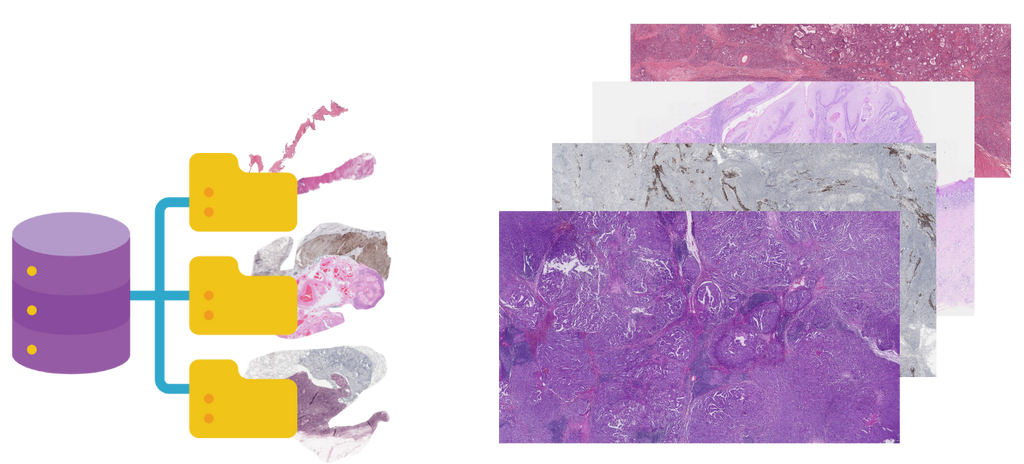
The goal of this project is to push the boundaries of self-supervised training in digital pathology by increasing the training set size by a factor ten compared with the state of the art, using both digital archival material from the Radboudumc, as well as a broad collection of publicly available data from grand challenges available via the grand-challenge.org platform.
Furthermore, we will investigate the benefits of learning a general-purpose representation of pathology images beyond H&E, also including immunohistochemical staining. The goal is to build some form of general-purpose pan-cancer histopathology encoder that can be used as a backbone to improve downstream tasks such as image classification, detection or segmentation. This project is intended to be a cross-application project and the resulting model will be tested on different clinical tasks.
Solution
Recent approaches like SimCLR[1], DINO[2], BYOL[3] and other similar self-supervised methods have proven their utility in learning good representations of histopathological images, where the amount of data in a single slide is very large, given the size of gigapixel images used in pathology. However, recent work in this field has solely paired self-supervised approaches with publicly available datasets such as TCGA, which contains 11,000 whole-slide images, solely stained with standard haematoxylin and eosin staining (H&E). Pushing the boundaries of the data set size might unlock the possibility of learning a very high-level representation of the data that can improve performance of state-of-the-art downstream tasks, such as image segmentation, classification, or object detection. Several AI tasks will be selected as demonstrators to test the model’s performance in different settings.
[1] Chen et al, A Simple Framework for Contrastive Learning of Visual Representations
[2] Caron et al, Emerging Properties in Self-Supervised Vision Transformers
[3] Grill et al, Bootstrap your own latent: A new approach to self-supervised Learning
Data
In this project we intend to use the digital archival material from the Radboudumc, as well as a broad collections of publicly available data from grand challenges available via the grand-challerge.org platform.
Approach
The student will be supervised by a research member of the Diagnostic Image Analysis Group and Computational Pathology Group whose research is dedicated to analyses of histopathological slides with deep learning techniques. The student will have access to a large GPU cluster.
Requirements
The candidate should have: - a major in computer science, biomedical engineering, artificial intelligence, physics, or a related area in the final stage of master level studies are invited to apply; - affinity with programming in Python; - interest in deep learning and medical image analysis.
Information
- Project duration: 6 months
- Location: Radboud University Medical Center
- For more information, please contact Marina D’Amato

